GCSE Biology - AQA
1.1.4 - Advances in Microscope Technology
Jump to:
Advances in Microscope Technology
Microscope technology has developed greatly since light microscopes were first invented hundreds of years ago. As microscope technology has improved, biologists have been able to learn more and more about cells and sub-cellular structures.
The invention of more powerful lenses for light microscopes
One advancement in microscope technology has been the invention of more powerful lenses for light microscopes.
More powerful lenses give greater magnifications, making it possible to see the cell's sub-cellular structures in more detail.
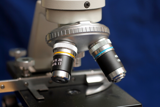
The invention of more powerful lenses has enabled light microscopes to achieve greater magnifications. (Photo by Krzysztof (Kriss) Szkurlatowski from FreeImages)
The invention of stains
Another major advancement has been the development of stains.
Stains are coloured chemicals which bind to specific sub-cellular structures when they are added to cells.
For example, there are some stains that bind to the nucleus and other stains that bind to cell membranes.
Most sub-cellular structures are transparent, making them difficult to see. Because stains are coloured and each one only binds to certain structures, they make it much easier for biologists to view the sub-cellular structures they are interested in.
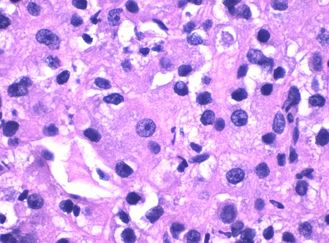
Human cancer cells stained with one stain that turns the cytoplasm pink and another that turns the nuclei blue. Viewed through a light microscope.
The invention of electron microscopes
In the 1930s, a new type of microscope called an electron microscope was invented.
Instead of using rays of light, an electron microscope creates an image by firing a beam of electrons at the sample.
An electron microscope has a much higher magnification and resolving power than a light microscope.
Electron microscopes have lead to great increases in our understanding of cells, because they make it possible to view cells in much finer detail, allowing biologists to see and understand many more sub-cellular structures.
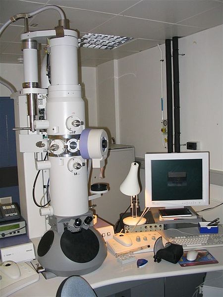
An electron microscope (Photo: Electron Microscope by David J Morgan from Wikimedia Commons - licensed under CC BY-SA 2.0)
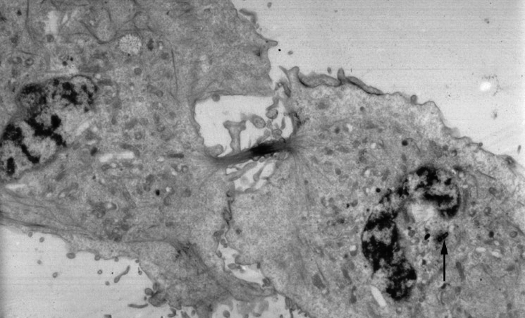
Image captured with an electron microscope of a cell dividing in two.
Flashcards
Flashcards help you memorise information quickly. Copy each question onto its own flashcard and then write the answer on the other side. Testing yourself on these regularly will enable you to learn much more quickly than just reading and making notes.
1/5
How has microscope technology improved over time?
2/5
What is a stain?
3/5
What is an electron microscope?
4/5
What are the advantages of electron microscopes over light microscopes?
5/5
How have electron microscopes helped to advance cell biology?
Donate
Please consider donating to support Mooramo. I am one person doing this whole project on my own - including building the site, writing the content, creating illustrations and making revision resources. By making a one-time or repeating donation you will buy me time to work on Mooramo, meaning that I can get new content on here more quickly.
Donate