GCSE Biology - AQA
1.1.3 - Microscopes
Jump to:
Microscopes
A microscope is a device used to view things that are small, such as cells and sub-cellular structures. The microscope creates an enlarged image of whatever is placed in it, making it possible to study things that would otherwise be too small to see.
There are several different types of microscopes, each with their own advantages and disadvantages.
Light microscopes
The first type of microscope to be invented was the light microscope. A light microscope works by shining light through the sample. This light then passes through two lenses, which magnify the image, before passing into the eyes of the person using the microscope.
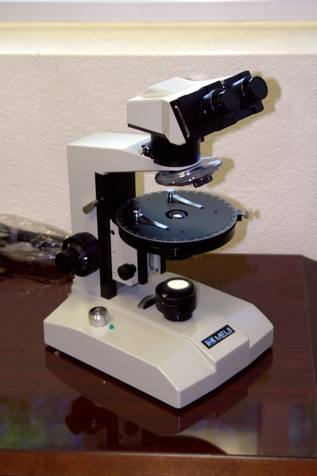
A light microscope. (Photo by dustin day from FreeImages)
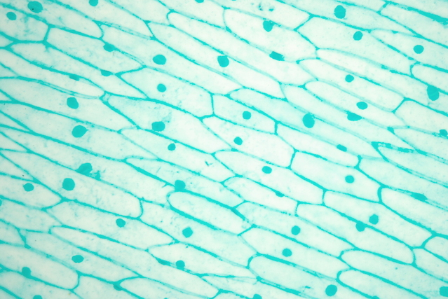
Cells from a leaf of a garlic plant, viewed through a light microscope. (Photo by Krzysztof (Kriss) Szkurlatowski from FreeImages)
In order to observe cells using a light microscope, the cells first need to be placed on a thin piece of glass called a slide.
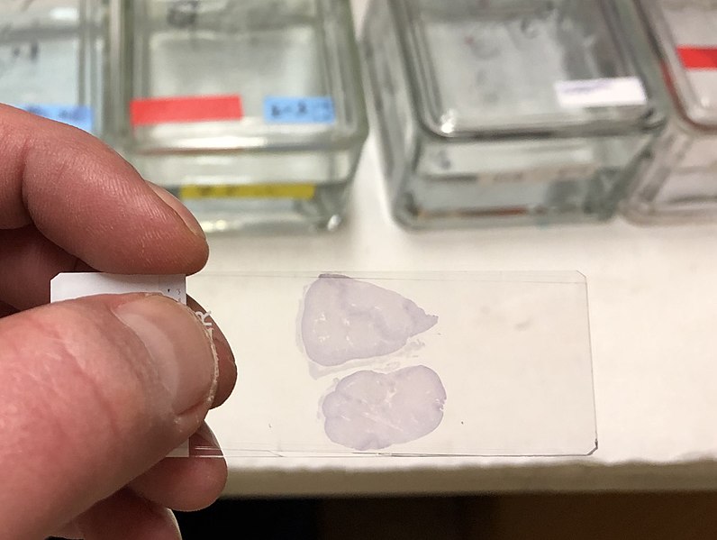
A microscope slide with a sample on it. (Photo: Stained microscope slide by Waughd on Wikimedia Commons - licensed under CC BY-SA 4.0)
The slide is then placed on the microscope and light shines through it, allowing a magnified image of the cells to be seen through the microscope's eyepiece.
Magnification
The magnification of an image is the number of times larger the objects in the image are than the actual objects.
For example, if you are looking at a cell through a microscope and the width of the cell in the image is two hundred times greater than the width of the actual cell, then you would say that the image has a magnification of 200X (pronouned '200 times').
The magnification of a light microscope can be changed by using different lenses.
The maximum amount of magnification possible is different for different types of microscopes.

Magnification means that the object in the image is bigger than the actual object. In this case, the image has a magnification of 10X, meaning that the cell in the image is 10 times wider than the actual cell.
Resolution
Resolution, also known as resolving power, is the ability to distinguish between close together points in an image.
The more resolving power a microscope has, the closer together two objects can be and still be seen as two distinct objects.
If the microscope does not have enough resolving power to distinguish between two close together objects, then they will appear blurred together into one object.

Resolution is the ability of a microscope to distinguish between objects that are close together.
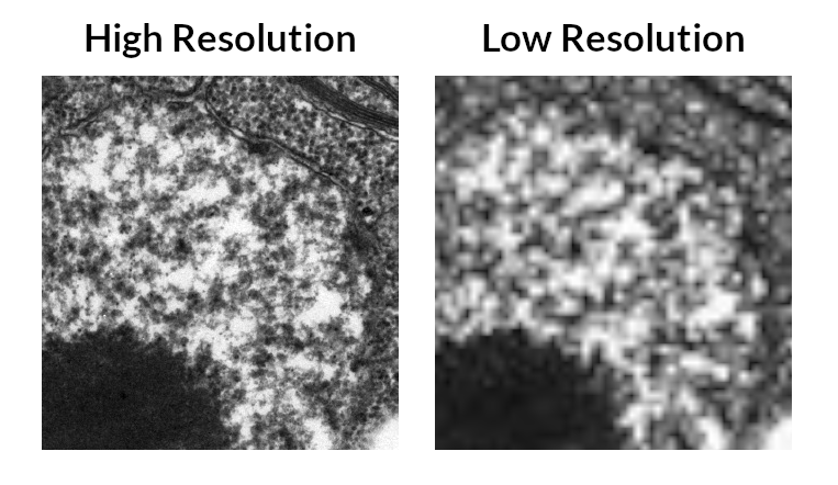
The same electron microscope image of part of a cell's nucleus shown at high resolution and low resolution (the nucleus is the round structure that takes up most of the image - not just the dark circle in the bottom left corner). The nucleus is surrounded by a double membrane - in the high resolution image this can be seen as two separate lines, whereas in the low resolution image it blurs into one line.
Flashcards
Flashcards help you memorise information quickly. Copy each question onto its own flashcard and then write the answer on the other side. Testing yourself on these regularly will enable you to learn much more quickly than just reading and making notes.
1/4
What is a microscope?
2/4
What was the first type of microscope to be invented?
3/4
What is magnification?
4/4
What is resolution?
Next Page
1.1.4 - Advances in Microscope Technology
Previous Page
1.1.2 - Scales and Sizes of Cells
Return to course page
Donate
Please consider donating to support Mooramo. I am one person doing this whole project on my own - including building the site, writing the content, creating illustrations and making revision resources. By making a one-time or repeating donation you will buy me time to work on Mooramo, meaning that I can get new content on here more quickly.
Donate